Investigation of patients with acute ankle ligament injuries is basically diagnostic imaging of various sorts. Other investigations may be indicated on a case by case basis.
Plain radiographs
The standard plain films of the ankle are mortise and lateral views. Radiography is not indicated in all acute ankle injuries. The Ottawa Ankle Rules have been proven to reduce unnecessary radiography while ensuring a high level of recognition of fractures. As well as malleolar fractures, plain films may show osteochondral injuries of the talus, lateral and posterior talar process fractures, avulsions of the superior peroneal retinaculum and calcaneal fractures. However, plain films will not generally demonstrate ankle ligament tears or instability.
In patients with a chronic history which is mainly of instability, radiographs are not routinely required. Where pain is a significant feature, plain AP and lateral radiographs of the ankle are generally obtained to evaluate the joint and look for associated injuries. We obtain these films standing to get an accurate assessment of joint space and alignment. However, plain films will not show 50% of osteochondral injuries, or soft tissue injuries such as peroneal tendon tears. No studies have examined the diagnostic yield of plain films in the chronically unstable ankle.
In patients with cavus or cavovarus feet we also obtain a standing hindfoot alignment view (Salzmann and El-Khoury 1995), which differentiates between malalignment within and below the ankle. Standing lateral and dorsoplantar views of the foot allow further evaluation of the deformity. Oblique hindfoot views may be obtained in cases of suspected subtalar pathology or fracture of the anterior calcaneal process.
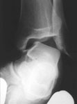
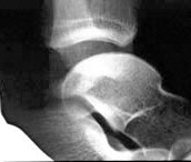
Ankle stress Xrays (L)anterior draw; (R)talar tilt
Stress radiography
Stress radiographs of the ankle have come to be considered the gold standard for the diagnosis of acute and chronic ligament injuries. An anteroposterior view with the hindfoot in maximal varus and a lateral with the hindfoot drawn anteriorly relative to the tibia are the standard views. Films of both ankles should be obtained for comparison. Frost and Amendola (1999) give a useful systematic review of stress radiography.
There is disagreement on many technical aspects of stress radiography. Stress radiography in the acutely injured ankle is painful. General and regional anaesthesia have been suggested to improve the accuracy of measurements. Becker et al (1993) showed that the anterior draw test gave greater values in both normal and injured ankles after peroneal nerve block, the difference being greater in the injured ankles. However their paper does not show whether this increased the diagnostic value of the test.
Ultrasound
In skilled hands, ultrasound has been shown to be technically capable of diagnosing acute ligament tears Campbell et al 1994). Van Dijk et al included ultrasound among the diagnostic modalities they studied and found it to be less accurate than clinical examination. In addition, the move away from acute ligament reconstruction has rendered the information from ultrasonography of the acutely injured ankle less relevant to the care of the patient.
MR
Magnetic resonance imaging allows evaluation of the ankle ligaments and also the tendons and bones. Studies in acutely injured ankles (Frey et al (1996), Magee and Hinson (1998), Tochigi et al (1998)), show associated injuries to the talus, subtalar ligament injuries and tears of the tibialis posterior and deltoid injuries, and some of these were associated with poorer outcome. In patients scheduled for surgical stabilisation of chronically unstable ankles, Chandnani et al (1994) showed MR arthrography to be superior to MR imaging and stress radiography for the detection of ATFL and CFL tears. MR is superior to plain radiography for the diagnosis and evaluation of osteochondral lesions of the talus (Hepple et al 2001). However, there have been few studies of how MR alters decision-making. In addition, MR cannot demonstrate instability without dynamic images.
We use MR:
- in patients with persistent severe pain after an acute injury, usually 4-8 weeks after injury. these patients may have intra-articular injuries which are amenable to arthroscopic treatment (Hepple et al 2004)
- in patients who fail rehabilitation for a chronic injury where pain in the joint (rather than impingement pain) is a significant problem, or where injection of an apparent impingement lesion is unsuccessful. the goal of the test is to look for an osteochondral lesion to support a decision on whether to offer an arthroscopy, and to guide visualisation at arthroscopy in what can be a tight ankle.