
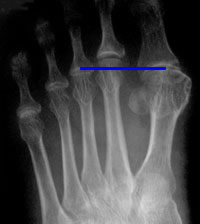
Long 2nd metatarsal (about 6mm beyond Coughlin's reference line) and slight joint distention
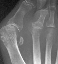
Dislocated 2nd MTP joint - note double shadow
Plain radiography
MTP instability is probably associated with a long second toe. Coughlin (1993) found that the 2nd metatarsal projected beyond the line between the 1st and 3rd metatarsals by an average of 9mm.
In early synovitis there may be slight distention of the joint. Plain films will also show dislocation and malalignment (much more often varus than valgus).
Arthrography, MR and ultrasound
Both MR (Yao 1994) and ultrasound (Gregg et al 2006) are capable of demonstrating plantar plate tears. Gregg's correlation with surgical findings suggests ultrasound may be more accurate, but the numbers were small. Interestingly, Gregg found plantar plate tears in 35% of asymptomatic feet. She alluded to the presence of partial thickness tears, which are poorly shown by MRI but well seen on ultrasound, and whose treatment is not yet defined.
Among others, Yao and Powless and Elze (2001) studied standard arthrography. Powless and Elze classified their findings on arthrography into type 1 (plantar extravasation indicating a plantar plate tear), type 2 (medial or lateral extravasation indicating a collateral ligament tear) and type 3 (combined tear) with some subclassification. They found plantar plate tears in 24 (41%), collateral tears in 19 (33%) and combined lesions in 15 (26%).
MR arthrography offers improved delineation of soft-tissue abnormalities, osteochondral lesions and bone oedema, but at higher cost and lower accessibility. Fluid can be injected under ultrasound control and extravasation observed to indicate a tear; this suggests a possible simple accessible test. Both ultrasound and MR can show plantar plate tears in which no extravasation occurs on injection, implying partial thickness tears.
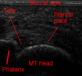
Ultrasound of complete plantar plate tear
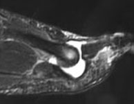
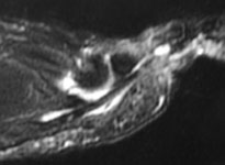
MR arthrogram shows contrast extravasation into the flexor tendon sheath