
There are two main classifications of Charcot neuroarthropathy.
Eichenholtz classification: disease progress
The Eichenholtz classification describes the evolution of the condition through time:
- Stage 1 – destruction
- Stage 2 – coalescence
- Stage 3 – consolidation
A "stage 0" has come into use to describe the swollen, hot, usually somewhat painful foot in which plain Xrays are normal. MR, however, shows bone oedema and stress fractures.
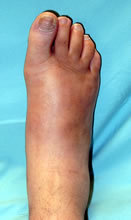
"Stage 0" - hot foot, normal Xrays. MR shows bone oedema and fractures
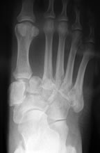
Stage 1 - fragmentation, bone resorption, dislocations, fractures
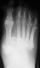
Stage 2 - coalescence, sclerosis, fracture healing, debris resorption
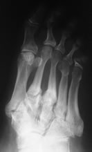
Stage 3 - remodelling
A new approach to disease staging?
The Eichenholtz classification is not entirely clear and its reproducibility unknown. Attention is increasingly focused on identifying Charcot arthropathy at a stage before bone destruction occurs (the so-called stage 0). At this stage bone damage can be identified by MRI or isotope scanning but not plain radiography.
Chantelau (2015) has proposed a new staging approach based on
- disease activity as demonstrated by MRI - Active (A) or inactive (B) disease
- presence of deformity - present (1) or absent (0)
- Active disease without deformity (A0) is the equivalent of "stage 0". This is the desirable stage for disease identification as treatment has a better chance of achieving healing without deformity and therefore with a lower long-term risk of ulceration and amputation
- Inactive disease without deformity (B0) iis the desirable end result of care
- Active disease with deformity (A1) encompasses Eichenholtz stages 1-3. Deformity and complications cannot be prevented but may be minimalised with appropraite treatment. Surgery in this stage would normally be avoided, although there are arguments to the contrary (Simon 2000, Salzman 2005).
- Inactive disease with deformity (B1) represents a stable end stage, but with an enhanced risk of ulceration. Orthotic treatment and follow-up is essential to minimise the risk. Surgery may be considered to reduce the risk of ulceration.
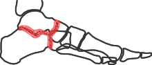
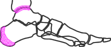
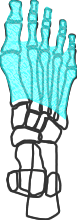
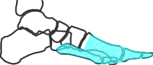
Brodsky classification: disease distribution
Charcot arthropathy usually begins in the tarsometatarsal region, but sometimes it is seen in the midtarsal or ankle joints, or as pathological calcaneal fractures. The distribution is usually expressed by the Brodsky classification. Schon has described a more precise classification, which has been validated, but it has not yet been shown that the additional complexity is clinically useful.

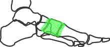
Type 1 (tarsometatarsal and lesser tarsus)
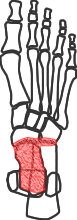
Type 2 (peritalar)

Type 3a (ankle) and 3b (posterior calcaneum)
Trepman added type 4 (multiple sites) and type 5 (forefoot) as shown here